AML is a cancer of myeloid progenitor cells. Changes in these cells stop myeloid blasts from becoming mature blood cells. As a result, there is a buildup of blasts in the marrow and blood. In turn, there are not enough red blood cells, platelets, and mature granulocytes.
To be diagnosed with AML, 20 percent (20%) or more myeloblasts must be present in the marrow or blood. In certain cases, a diagnosis of AML is possible with less than 20% blasts.
Types of AML
There are many types of AML. They are grouped and treated based on gene mutations and other features. Genetic and biomarker testing is used to identify the AML subtype. It is a standard part of testing at diagnosis.
- Acute myeloid leukemia (AML)
- Acute promyelocytic leukemia (APL)
- Blastic plasmacytoid dendritic cell neoplasm (BPDCN)
Tests for Acute myeloid leukemia
A diagnosis of AML is based on the presence of 20 percent (20%) or more myeloid blasts in the marrow or blood. This means that at least 2 out of every 10 marrow cells are blasts. However, in some cases a diagnosis of AML is possible with less than 20% blasts.
- Medical history and physical exam
- Complete blood count, platelets, differential, comprehensive metabolic panel, uric acid,lactate dehydrogenase, B12, and folic acid
- Blood clotting tests: PT, PTT, and fibrinogen
- Bone marrow core biopsy and aspirate with immunophenotyping
- Cytogenetic analysis (karyotype and FISH)
- Molecular tests of these and other mutations: ASXL1, c-KIT, FLT3 (ITD and TKD), NPM1, CEBPA, IDH1, IDH2, RUNX1, and TP53
- Comprehensive pathology report with genetics, blast count
- HLA typing for possible stem cell transplant candidates
- Brain CT without contrast, if central nervous system bleed suspected
- Brain MRI with contrast, if leukemic meningitis suspected
- PET/CT if extramedullary disease suspected
- Lumbar punctureonly if central nervous system involvement is suspected
HLA typing
Human leukocyte antigens (HLAs) are proteins found on the surface of most cells. They play an important role in your body’s immune response. HLAs are unique to each person.
They mark your body’s cells. Your body detects these markers to tell which cells are yours. In other words, all your cells have the same set of HLAs. Each person’s set of HLAs is called the HLA type or tissue type.
HLA typing is a blood test that detects a person’s HLA type. This test is done before a donor (allogeneic) blood stem cell transplant. To find a donor match, your proteins will be compared to the donor’s proteins to see how many proteins are the same. A very good match is needed for a transplant to be a treatment option. Otherwise, your body will reject the donor cells or the donor cells will react against your body. Blood samples from you and your blood relatives will be tested first.
Who needs HLA typing?
HLA typing should be done in all patients with newly diagnosed AML for whom allogeneic (donor) blood stem cell transplant is an option. HLA typing of family members up to 80 years of age is recommended. Tissue typing should include searches for other possible donors.
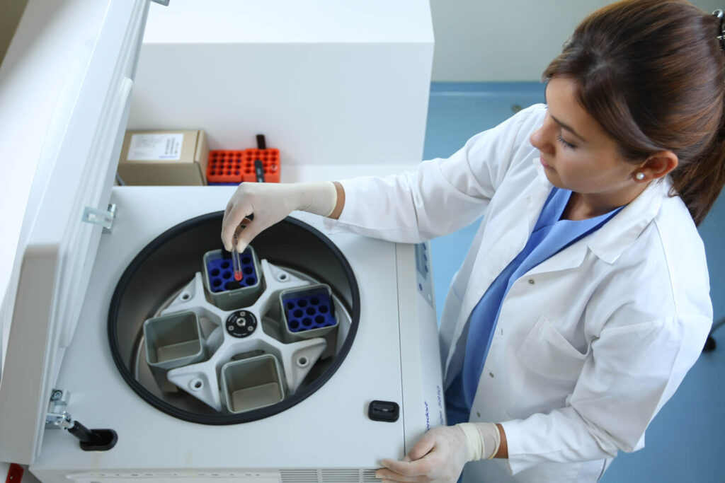
Bone marrow tests
Leukemia starts in the bone marrow. To diagnose AML samples of bone marrow must be removed and tested before starting any treatment. Your sample should be reviewed by a hematolog or hematopatolog who is an expert in the diagnosis of AML. Immunuphenotyping by flow cytometry of the bone marrow aspiration sample also is done and compared with morphology of the blasts.
Genetic tests
Genetic tests are used to learn more about your type of AML, to target treatment, and to determine the likely path your cancer will take (prognosis). This testing is different from family history genetic testing. AML genetic testing looks for changes only in the leukemia cells that have developed over time, and not changes in the rest of your body’s cells.
Inside our cells are deoxyribonucleic acid (DNA) molecules. These molecules are tightly packaged into what is called a chromosome. Chromosomes contain most of the genetic information in a cell. Normal human cells contain 23 pairs of chromosomes for a total of 46 chromosomes. Each chromosome contains thousands of genes. Genes tell cells what to do and what to become. Most genes contain information about a specific protein.
AML cells sometimes have changes in genes and chromosomes that can be seen under a microscope or found with other tests. Cytogenetic tests look for these changes or abnormalities.A karyotype and FISH are two types of cytogenetic tests used in AML.
Imaging tests
Imaging tests take pictures of the insides of your body. These tests are used to show which sites have leukemia. They can also show areas of infection or bleeding, which may impact your care.
Leukemia can spread outside the bloodstream to lymph nodes, liver, spleen, skin, and other organs. It rarely spreads to the lining of the brain and spinal cord.
A radiologist, who is an expert that looks at test images, will review your images and write a report.
Lumbar puncture
Leukemia can travel to the fluid that surrounds the spine or brain. This may cause symptoms. In order to know if leukemia cells are in your spinal fluid, a sample must be taken and tested to rule out a central nervous system disease.
A lumbar puncture is a procedure that removes spinal fluid. It is also called a spinal tap. A lumbar puncture may also be used to inject cancer drugs into spinal fluid. This is called intrathecal chemotherapy.
During a spinal tap, you will be lying down or sitting on an exam table. If lying down, your knees must be tucked up near your chest.
If sitting, you must lean forward toward your knees. The lower part of your back over your spine will be numbed. Next, a thin needle will be inserted between your spinal bones. You may feel some pressure. After the sample is taken, it will be sent to a lab for testing.