Acute lymphoblastic leukemia (ALL) is a fast- growing blood cancer that starts in disease- fighting lymphocytes of your immune system. In ALL, bone marrow makes too many immature lymphocytes called lymphoblasts. Lymphoblasts can crowd out other blood cells causing blood to not work as it should. Acute leukemias grow faster than chronic leukemias.
ALL most often affects B cells or T cells.
To be diagnosed with ALL, 20 percent (20%) or more lymphoblasts must be present in the bone marrow. This means that at least 2 out of every 10 marrow cells are lymphoblasts. In certain cases, a diagnosis of ALL is possible with less than 20% lymphoblasts.ALL can be found in bone marrow, blood, and organs such as the testicles or the central nervous system.
Types of ALL
ALL is a group of diseases. They are grouped and treated based on gene mutations and other features. Genetic testing is very important to identify the ALL subtype. It is a standard part of testing at diagnosis.
There are 2 types of ALL:
- B cell or B-ALL
- T cell or T-ALL
Within each type there are several subtypes, which are based mainly on:
- The type of lymphocyte (most often B cell or T cell) the leukemia cells come from and how mature the cells are. This is known as the immunophenotype of the leukemia.
- If the leukemia cells have certain gene or chromosome changes
Testing for ALL
Accurate testing is needed to diagnose and treat ALL. Test results will determine your treatment plan
- Complete blood count (CBC), differential, chemistry profile, liver function tests (LFTs)
- Tumor lysis syndrome (TLS) panel: LDH, uric acid, potassium (K), calcium (Ca), phosphorus (Phos)
- Disseminated intravascular coagulation (DIC) panel: d-dimer, fibrinogen, prothrombin time (PT), and partial thromboplastin time (PTT)
- Testing for hepatitis B and C, HIV, cytomegalovirus (CMV) antibodies (Ab) Pregnancy testing, fertility counseling, and preservation
- CT and MRI of head with contrast, if the patient has neurologic symptoms. Lumbar puncture (LP) with intrathecal (IT) chemotherapyalso can be necessary.
- CT of neck, chest, abdomen, pelvis with contrast. PET/CT is possible Testicular exam, including scrotal ultrasound as needed
- Screen for opportunistic infections, as needed
- Echocardiogram or cardiac nuclear medicine scan should be considered in all patients
- Strongly consider early transplant evaluation and donor search
Lumbar puncture
A lumbar puncture (LP) or spinal tap is a procedure that removes spinal fluid. It is also used to inject chemotherapy into the spinal fluid. This is called intrathecal (IT) chemotherapy.
At the time of IT, a sample of your spinal fluid will be taken and tested. Leukemia can travel to the cerebrospinal fluid (CSF) that surrounds the spine or brain. This is called central nervous system (CNS) disease. Systemic therapy and IT therapy are given together to prevent CNS disease. This is called CNS prophylaxis. IT therapy needs to be given even if the CNS is negative because leukemia is likely to return here.
Bone marrow tests
Leukemia starts in the bone marrow. To diagnose ALL, samples of bone marrow must be removed. Lab results will be used to confirm the disease. Your bone marrow will also be tested to see how well treatment is working.
There are 2 types of bone marrow tests that are often done at the same time:
- Bone marrow aspirate
- Bone marrow biopsy
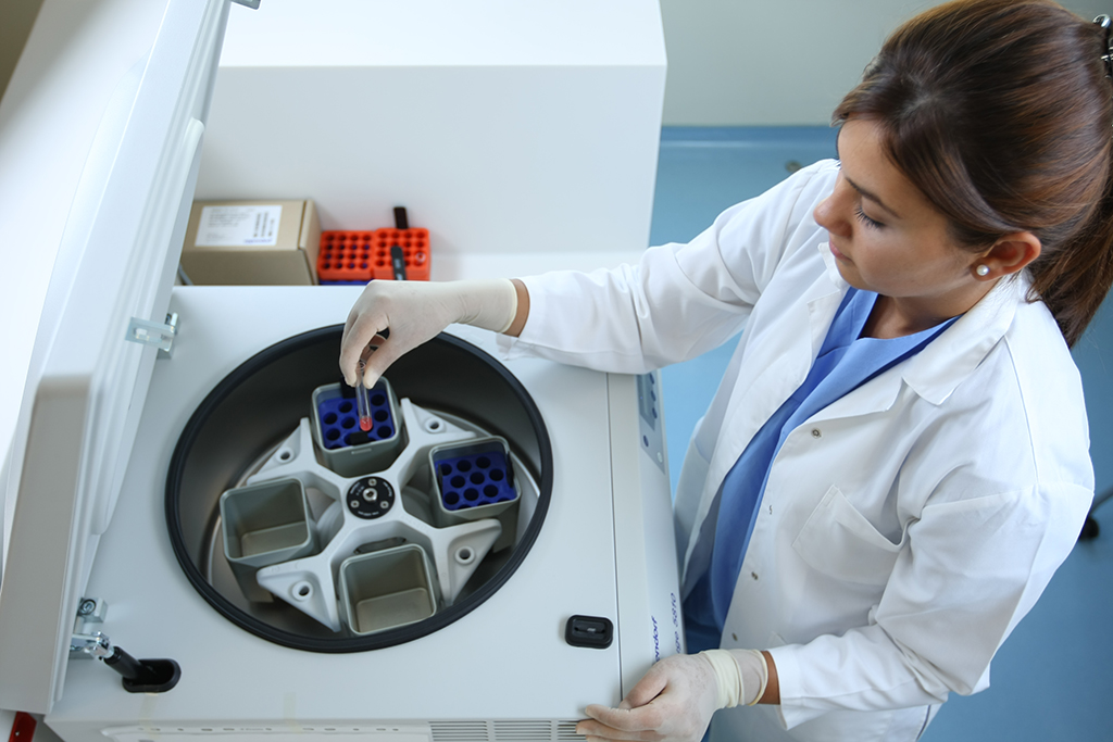
Your bone marrow is like a sponge holding liquid and cells. An aspirate takes some of the liquid and cells out of the sponge, and a biopsy takes a piece of the sponge. The samples are usually taken from the back of the hip bone (pelvis). You will likely lie on your belly or side. Your doctors will first clean and give sedation or numb your skin and outer surface of your bone. For an aspirate, a hollow needle will be pushed through your skin and into the bone. Liquid bone marrow will then be drawn into a syringe. For the biopsy, a wider needle will be used to remove a core sample.
Flow cytometry
Flow cytometry is a laboratory method used to detect, identify, and count specific cells. Flow cytometry involves adding a light-sensitive dye to cells. The dyed cells are passed through a beam of light in a machine. The machine measures the number of cells, things like the size and shape of the cells, and proteins on the surface of thousands of cells.Flow cytometry may be used on cells from circulating (peripheral) blood, bone marrow, or a biopsy. The most common use of flow cytometry is in the identification of markers on cells (called immunophenotyping). This test is important because the immunophenotype put the differentiate B-ALL or T-ALL.Certain biomarkers such as CD19, CD20, and CD22 are targeted in ALL treatment.
Based on immunophenotype, ALL can be placed into 2 general groups:
- B-cell ALL(CD19, CD79a positive)
- T-cell ALL(CD2, CD7,CD 3 positive)
B-ALL
B-ALL is the most common type of ALL. It starts in immature cells (lymphoblasts) that would normally develop into B lymphocytes. Subtypes include early precursor B cell (early pre-B cell) and pre-B cell. Mature B-cell ALL (Burkitt lymphoma) is a rare subtype. B-ALL can be Philadelphia chromosome-positive (Ph+) or Philadelphia chromosome-negative (Ph-). Another B cell ALL type is Philadelphia like B-ALL.
T-ALL
T-ALL starts in lymphoblasts that would normally develop into T-cell lymphocytes.This type is less common, and it occurs more often in adolescent boys. A protein called CD3 is typically found in T-ALL. Others may be present. T-ALL does not have the Philadelphia chromosome . Early T-cell precursor is a distinct subtype of T-ALL.
Genetic tests
Genetic tests are used to learn more about your type of ALL, to target treatment, and to determine the likely path your cancer will take (prognosis).Inside our cells are deoxyribonucleic acid (DNA) molecules. These molecules are tightly packaged into what is called a chromosome. Chromosomes contain most of the genetic information in a cell. Normal human cells contain 23 pairs of chromosomes for a total of 46 chromosomes. Each chromosome contains thousands of genes. Genes tell cells what to do and what to become.ALL can cause changes in genes and chromosomes in blood cells. Genetic tests look for these changes or abnormalities. You may be placed into a risk group based on the types of genetic abnormalities found.
Next-generation sequencing
Next-generation sequencing (NGS) is a high- throughput method used to determine a portion of a person’s DNA sequence.
FISH
Fluorescence in situ hybridization (FISH) is a method that involves special dyes called probes that attach to pieces of DNA. For example, the probes attach to the BCR gene and the ABL gene. The BCR-ABL1 geneis detected when the colors of the probes overlap by translocation. A translocation is the switching of parts between two chromosomes. Another possible translocation found in ALL is KMT2A or t(v;11q23.3). FISH can look for other translocations that are too small to be seen with other methods.Since this test doesn’t need growing cells, it can be performed on either a bone marrow or blood sample. However, FISH can only be used for known changes. It cannot detect all the possible changes found with a karyotype.Sometimes, a bone marrow sample will still be needed to get all of the information your doctor needs to help plan your care.
Karyotype
A karyotype is a picture of chromosomes. Doctors look for whether 46 chromosomes or 23 pairs are present. They also look for extra, missing, or abnormal pieces of chromosomes, such as the BCR-ABL1 gene. Since a karyotype requires growing cells, a sample of bone marrow must be used.
PCR
A polymerase chain reaction (PCR) is a lab process that can make millions or billions of copies of your DNA (genetic information). PCR is very sensitive. It can find 1 leukemia cell among more than 100,000 normal cells.A special PCR called quantitative reverse transcriptase-polymerase chain reaction (qPCR) measures the number of cells with the BCR-ABL1 gene. This test might be referred to as real-time or reverse transcriptase (RT) PCR.
Philadelphia chromosome All cells in our body contain genetic information organized in chromosomes. A cell must make a copy of its chromosomes before dividing into two cells. Sometimes, there are mistakes in the copies. One type of mistake is when parts of two chromosomes break off and switch with each other. This is called a translocation. It can result in a fusion gene. There are a few different translocations that might be found in ALL. One is the Philadelphia chromosome (Ph).In the Philadelphia chromosome, a piece of chromosome 9 and a piece of chromosome 22 break off and trade places with each other. These pieces then fuse together on chromosome 22. This new, abnormal chromosome 22 is referred to as the Philadelphia chromosome. You might see it written as Ph-positive (Ph+).The piece of chromosome 9 is a gene called ABL. The piece of chromosome 22 is a gene called BCR. When these genes fuse together on chromosome 22, the BCR-ABL1 gene is formed. BCR-ABL1 is a fusion gene. It is not found in normal blood cells. It is not passed down from parents to children.BCR-ABL1 makes a new protein that leads to uncontrolled cell growth. Treatment for Ph+ ALL aims to stop the activity of the BCR-ABL fusion protein.